
The use of time‐resolved fluorescence has expanded as the relative cost of instrumentation has decreased in recent years. One area where this is especially true is in the food industry, where time‐resolved fluorescence has been applied in the characterisation of food stuffs as well as aspects related to food safety and degradation.
Fluorescence in food
There are a variety of fluorescent substances in food stuffs, ranging from amino acids (Phe,Tyr,Trp) in proteins to vitamins to chlorophyll, for example. Fluorescent substances are therefore contained in both plant and animal derived foods, which means that fluorescence techniques can be widely applied. The principal area of usage includes food safety and storage. The monitoring of contaminants, such as
bacteria, toxins and insects, is possible using fluorescence and the technique can also be applied to the development of novel foodstuffs. The versatility of fluorescence is evident from its usage with dairy products, vegetable oils, beers /wines, animal proteins, enzymes and food colourings to name just a few. Although intrinsic fluorophores can be used, at times it can be advantageous to add an extrinsic probe.
In this note a couple of examples will be given, looking at the use of extrinsic probes in food related micro‐emulsions and intrinsic fluorescence in a staple foodstuff elucidated using time‐resolved fluorescence.
Oil in water microemulsions
Microemulsions have recently been applied to stabilise food colourings and deliver bioactives, as well as being used in low fat spreads. However, there are naturally occurring microemulsions such as milk. There are many dyes available which can either partition to the aqueous or oil phase, or to specific constituents in the emulsion. Fig. 1, recorded with the CCD camera on a HORIBA Scientific DynaMyc, is an example of a solution of non dairy creamer with rhodamine B and nile red used as extrinsic dyes. The field of view is approx. 1.3 x 1mm.
Part of the reason to study these systems is to ascertain their stability by characterizing their make up. Information concerning the membrane structure can be obtained from how the probe molecule locates, ie if there is the formation of lipid rafts or other inhomogeneities. Fig. 2 shows an uneven location of the fluorophore in an oil droplet from a creamer indicating a non homogeneous structure.
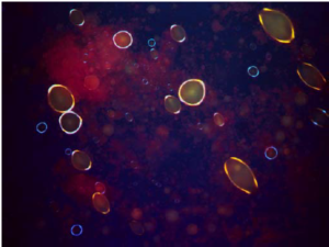
Fig. 1 Camera image using white light illumination and filtercubes with 500nm excitation, >515nm emission.
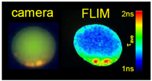
Fig. 2 Fluorescence camera and FLIM image of a stained fat droplet (~15m in diameter) recorded using a DynaMyc.
Learn More?
Please click on ‘Request Application Note’ and download the full application note ‘Solid Time-resolved fluorescence for monitoring food composition’.