Generating three-dimensional models of biological tissues
Using a TM4000plus SEM with Hitachi 3D map software
In conventional biomedical research, biological tissues are examined with light microscopy. This modality is easy to use and provides magnified images of the samples, but three-dimensional (3D) information is missing. Electron microscopes provide high resolution images with a larger field of depth, which creates a typical 3D appearance. Recently, Sawaguchi et al. (2018) developed a survey for assessing tissue architectures with low-vacuum SEMs. This application note shows how an easy to handle tabletop SEM, the TM4000Plus from Hitachi, generates 3D models of biological tissues.
Sawaguchi et al. analyzed cells from different organs obtained from rats using a light microscope and a TM4000Plus SEM. For oncological research, tumor cells are cultured and a morphological comparison is made between in vivo xenografts and in vitro grown 3D tissue.
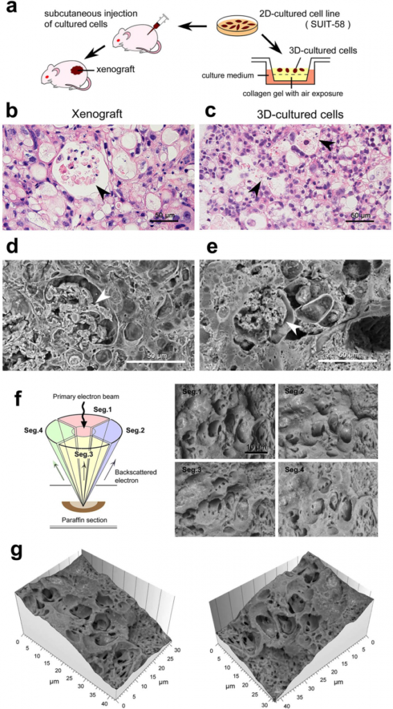
Cultured 2D cells are separated in two groups: one part is injected subcutaneously as a xenograft and one part is cultured in a medium to a 3D tissue (A). Results are obtained with a light microscope (B&C) and a Hitachi low-vacuum SEM (D&E). In both type of microscopes, vacuole formation enclosing cell debris is observed and indicated with arrow heads. The strength of SEM is demonstrated in the consecutive pictures F & G: with a four-segmented BSE detector, four images are collected simultaneously (F) and reconstructed to a 3D model of the tissue architecture (G). This application demonstrated how SEM remains superior to analyze tissue architectures. Read the full paper for more examples of 3D SEM analyses in biomedical applications.
This paper shows how electron microscopy literally adds an extra dimension to conventional microscopy. The Hitachi map 3D software offers many functionalities like profile and particle analysis, grain detection and image stitching and is available for many SEM models.
Reference:
Sawaguchi, A., Kamimura, T., Yamashita, A. et al. Informative three-dimensional survey of cell/tissue architectures in thick paraffin sections by simple low-vacuum scanning electron microscopy. Sci Rep 8, 7479 (2018) doi:10.1038/s41598-018-25840-8