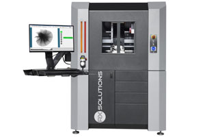
X-ray micro CT (microtomography)
X-ray micro CT (microtomography) uses the same technology as an ordinary CT (CAT) scan in a hospital.
X-ray micro CT (microtomography) uses the same technology as an ordinary CT (CAT) scan in a hospital.

♦ EasyTom S | High performance compact CT system
♦ EasyTom L | High resolution micro and nano CT system
♦ EasyTom XL | Industrial micro CT system
♦ UltraTom | Ultimate flexibility micro CT
X-ray micro CT (microtomography) has a resolution in the micrometer range, and can provide both high resolution and a large field of view.
Measured images are reconstructed by dedicated software. This technique allows you to see inside an object and look at it from all angles. The non-destructive technology means it is also possible to make a 4D time series of images, following any changes over time inside the sample.
ST Instruments offers instrumentation designed and produced by NeoScan. This company also provides its own all-in-one software tool for intuitive scanner control and processing data, from acquisition to 2D and 3D image reconstruction.
The technology uses an X-ray cone beam, which will pass through the sample to a detector. The sample volume which it penetrates will affect the detected signal. A 2D slice is made by rotating the sample (or the beam and detector) 180 to 360 degrees to make a series of projection images. These are then combined into a cross-sectional image via a computational process. The slices are then processed into a 3D model, the final image. This process is called computational tomography.
The resolution of the image is determined by the size of the x-ray focal spot, number of projection views per rotation, the detector cell size, and the software algorithms used in image reconstruction.
An X-ray micro CT gives you X-ray vision: it offers a non-destructive high resolution look inside your samples. It helps you to study the texture, detect faults, or measure porosity in for example bone or teeth, in pharmaceuticals (pills or capsules) and in materials or devices. By increasing the voltage of the electron beam, it is possible to penetrate more dense materials. Anything from leaves through polymers to metals can be imaged using X-ray micro CT.
The images will provide quantitative information that cannot be obtained by other techniques. As scanning is non-destructive, it is also possible to image the same object several times, to create a time sequence or measure the impact of a manipulation of the sample. Also, it is possible to subject a sample to additional analytical techniques after the micro CT scan.
Shale
Shale, a fine-grained sedimentary rock, is composed of highly compacted clay particles and contains nanometric porosity and silt-sized particles of other minerals. X-Ray micro CT has been used to study the shale structure at meso scale and observe the evolution of 3D crack networks in samples of shale under uniaxial compression at different levels of load. It also reveals the distribution of minerals and the morphology of breaks between the thin shale laminae.
Based on the acquired images, statistical morphological measures can be calculated, such as the volume fraction of cracks, and crack aperture spatial distribution. Furthermore, the technique can be used to image oil shale to analyse the nature of the pore network structure before and after the pyrolysis used to extract the oil.
Bone
X-ray micro CT is a standard tool in the evaluation of bone architecture. A scan of a bone at a pixel size of 8 µm, using the NeoScan S80 system non-destructively reveals the trabecular structure inside the bone in 3D. Such a scan is often faster than histology.
Micro CT scans also allow the study of the network of porous canals in cortical bone that facilitates the distribution of neurovascular structures throughout the cortex. Measuring the size, spacing, and volume of the canals helps to assess the mechanical properties of bone.
Electronics
As electronic equipment becomes ever smaller, there is a need to test and check components for deformation caused by mechanical or thermal loads. X-ray micro CT allows this in a non-destructive manner. It is for example possible to scan an entire smart watch of 50 x 42 x 13 mm at 18 µm pixel size, revealing all inner electronics.
Our technique can be used to detect failures in for example soldering in electronic circuits and allows the non-destructive evaluation of internal layers of multi-layered printed circuit boards with their components.