
Due to the numerous applications it presents, the microfluidics field has experienced enormous developments in the past few years. Lab-on-a-chip, organ-on-a-chip, point of care devices, cell capture, chemical and biological analysis are some of the examples for direct microfluidic applications. Regarding uses, microfluidic devices have different geometries which can be as complex as needed, but one of the basic structures that comprise these microfluidic device is the microchannel. Several materials have been reported for the fabrication of the microchannel, and the suitability of one or another depends on the fabrication technique. Some of these materials are polymers, silicon or glass. Examples of manufacturing techniques are soft-lithography, photolithography or thermofusing techniques. But when using soda lime glass as material which was chosen due to its robustness, chemical resistance, transparency and low cost- direct laser writing presents itself as one of the most suitable techniques. It is accurate and versatile, generating very complex geometries quickly. Moreover, due to its non-contact nature, there is no contaminant and does not require clean room facilities. When working in micron dimensions for this application, it is crucial to have a very good image of the topography to ensure its quality, as well as all the information about the channel dimensions. In this report, the structures manufactured by direct laser writing are fully characterized by means of confocal microscopy.
Measurements
Microchannels were fabricated over soda lime glass by direct laser writing. The laser employed was a Rofin Nd:YVO4 system, with 20 ns pulse duration and 1064 nm central wavelength. The setup was composed by a galvanometer system that addresses the beam and allows the fabrication of complex structures with no need of sample movement. The laser beam was focused over the substrate surface by using a lens of 100 mm focal length that ensures a working area of 80×80 mm2. Soda lime glass was obtained from a local supplier.
In order to obtain a proper aspect ratio of the structures, several laser scans were applied to the sample. Therefore, a study of the evolution of the topography was performed. Microchannels with scans from one to ten times were manufactured. Using Sensofar S neox 3D optical profiler, a confocal image of the structured area was captured using a 20X magnification objective. The surface profile was generated and the evolution of the depth of microchannels with the laser scans was depicted (Figure 1).
Roughness value of the microchannel walls are a critical value to know since it should be low enough for microfluidic applications. Thanks to Sensofar S neox 3D optical profiler and SensoMAP analysis software, it was possible to obtain roughness values from small areas. The topography of the bottom of a microchannel with eight laser scans, obtained with a 50X magnification objective, was selected for the study (Figure 2).
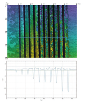
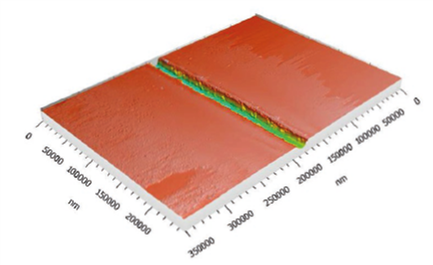
Figure [1] Confocal image of several microchannels with different laser scans, from one (left) to ten (right). The profile of the channels is also generated
Figure [2] 3D topography of one microchannel fabricated with laser and the roughness parameters of the bottom of the channel, according to ISO 25178
Learn More?
Please click on ‘Request Application Note’ and download the full application note ‘Characterization of micro-channels fabricated with laser for microfluidic applications’.