Surface topography is currently considered one of the most important surface properties regarding the biological response. Accordingly, an intense amount of implant research has been focused on the development of new surface treatments to increase surface roughness, aiming to enhance the biological response and ultimately, the osteointegration. However, the higher peaks and features of the enhanced roughness may break and detach or be modified during the insertion procedure into bone. Therefore, an important aspect to consider, though often overlooked, is whether the increasingly complex surface features found on modern dental implants are retained after insertion.
The aim of this research was to investigate the impact of the insertion procedure on the surface topography of dental implants. To achieve this, it is necessary to evaluate if the surgical procedure degrades the enhanced surface roughness by measuring and comparing the surface topography both before and after insertion exactly at the same locations. The present paper is a part of a research1,2 carried out by Dr. Alaitz Zabala, Dr. Wilson Tato, Dr. Andrea Aginagalde and Dr. Iñigo Llavori.
Measurements
Wennerberg et al.3 took the first steps in measurements’ standardization suggesting some guidelines for dental implant characterization: three samples in a batch must be evaluated, three dimensional measurements must be made at three evaluation areas (tops, flanks and valleys), the filter size must be specified, and at least one of each height, spatial, and hybrid parameters should be presented. In addition, Wennerberg et al. claimed that Confocal profilometers and Interferometers are the only acceptable methods to evaluate densely threaded oral implant designs. This is one of the main reasons why the commercial 3D optical profilometer Plμ (former version of S neox, Sensofar Metrology, Spain) was chosen to carry out this work. This unique optical profilometer is the only one in the market that includes Confocal, Interferometry (Phase Shifting and Coherence Scanning Interferometry) and Focus Variation techniques embedded into the same optical instrument. This feature allows for quickly evaluating dental implants with different optical techniques. Many international dental research labs have used Sensofar’s equipment for dental implants surface characterization, validating the technology and making it one of the gold standards for this purpose.4–12
In our study dental implants were measured with confocal at four different locations (Fig. 1), both before and after insertion in cow rib bone (which mimics human bone, Fig. 2).

Figure [1] Denomination of the different evaluation sites and profile examples
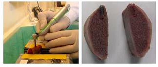
Figure [2] Insertion procedure in fresh caw rib bone under standard procedures. (a): insertion process; (b): non traumatic implant retrieval by separating the two previously generated bone block halves
Due to the complex geometry of threaded dental implants, the measurement of different locations of the implants and the location of the same region both before and after insertion present a challenge.
Different locations were correctly positioned by means of an ad-hoc developed dental implant handling and positioning device with 3 degrees of freedom (Fig. 3).
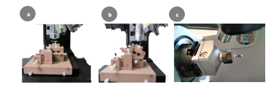
Figure [3]. Dental implant handling and positioning device for correct positioning of implants. (a): measurement of the top site; (b): measurement of the flank site; (c): both measurements can be done by a specific rotational system too (i.e. S neox Five Axis)
However, the most advanced Sensofar’s system includes a rotational module (i.e., S neox Five Axis) that enables an automated full three-dimensional surface characterization. Measurements were acquired using a 100X SLWD
magnification objective in order to acquire all topographical features in detail. The measurements were postprocessed by means of the metrology software SensoMAP (Digital Surf ) in order to remove form (second order polynomial fitting), noise (Gaussian filter cut-off 0.36 μm) and waviness (Robust Gaussian filter cutoff 50 μm). These values were considered optimal for this application given previous studies.1 Topographical parameters from ISO 25178-2 belonging to height, spatial, hybrid and functional families were calculated in order to fully characterize the surface properties.
Fig. 4 shows axonometric representative projections of the measurements taken at the same regions before and after insertion for the acid etched implants. The sites that showed statistically significant topographical parameter variations (p<0.05) are marked with *. It can be noted that the sharp peaks initially present became less prominent or had completely gone after insertion in all the sites under study except the neck.
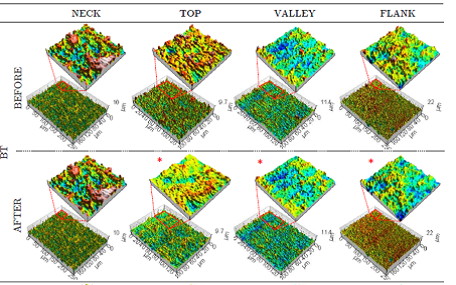
Figure [4] Representative axonometric projections of the measurements of an acid etched dental implant at the four evaluation sites both before and after insertion. Those sites presenting statistically significant topographical parameter variations are marked with*.
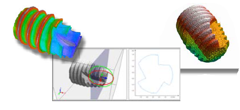
Figure [5].Different views from the measurement profile process.
Conclusion
Confocal technology has proven to be an effective technique to investigate and characterize surface topography and the damage generated in modern dental implant surfaces after the insertion into bone. This non-destructive measuring technique allows characterizing different locations of the complex threated dental implants with high resolution. Qualitative and quantitative analysis were done to evaluate the surface modifications. The following main points can be concluded from the various components of this study:
- Surface damage varied at different evaluation sites for each implant system (neck, top, valley and flank) and also along the implant within each site.
- The developed interfacial ratio (Sdr) variation correlated positively with the material volume variation (Vm) and is considered to be the best damage indicator.
- The use of functional volume parameters of ISO 25178 standard is suggested rather than the Rk family parameters defined in the European report EUR 15178N due to the higher sensitivity encountered.
- The 3D optical profilometer S neox (Sensofar, Spain) has been shown to be an accurate tool to investigate the surface topography of dental implants before and after insertion into bone. More advanced tools such as the S neox Five Axis, would not only enable faster and more automated high resolution measurements, but also the 3D reconstruction of the complete dental implant’s shape, which can be of interest for wear analyses or quality control in further studies.