Grain boundary embrittlement and high temperature grain boundary instability are important factors in the quest for higher performance metals for many industrial applications. Of particular importance is the composition of interphase boundaries (the interface between separate phases) in these metals. Scanning Auger spectroscopy combined with in-situ facture analysis provides a unique technique to expose the surfaces of the interphase boundaries and provide compositional analysis and elemental mapping of these surfaces. Shown below are secondary electron and Auger images that reveal the presence of multiple phases within the matrix of a ductile iron sample and exposed interphase boundaries.
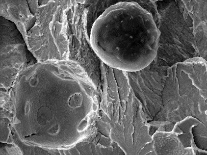
Fig. 1: Secondary Electron image
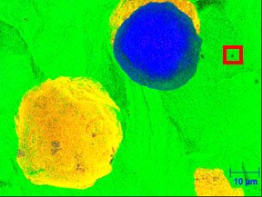
Fig. 2: Fe (green), C (blue), and Sn (yellow) Auger maps
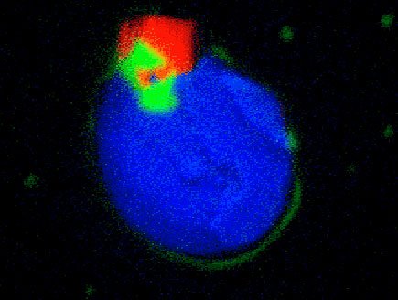
Fig. 3: Mg (green), Ca (blue), and Ti (red) Auger maps
The secondary electron image in figure 1 shows the microstructure of a ductile iron fracture surface including graphite nodules and craters where graphite nodules have fallen out as a result of the fracture. The AES maps in figure 2 show the ability to map across the graphite nodule and the crater where Sn has segregated to the nodule / iron interface. AES maps in figure 3 show the complex composition of a small precipitate observed in figure 2.